Choose a channel
Check out the different Progress in Mind content channels.

Progress in Mind

In all affected brain regions, α-synuclein accumulates only in specific cell types, and only in certain cells. Not all neurons are vulnerable to Lewy pathology; and not all glia accumulate α-synuclein. Factors determining the selective nature of neural degeneration are central to the better understanding of Parkinson’s disease and other synucleinopathies.
Some cells never accumulate α-synuclein though surrounding cells do. This is one of the paradoxes of Parkinson’s disease (PD). Even with end-stage disease, and even in the substantia nigra, not all neurons are affected by α-synuclein deposition. Across the brain, the proportion of targeted cells affected may be as low as 5-15%.
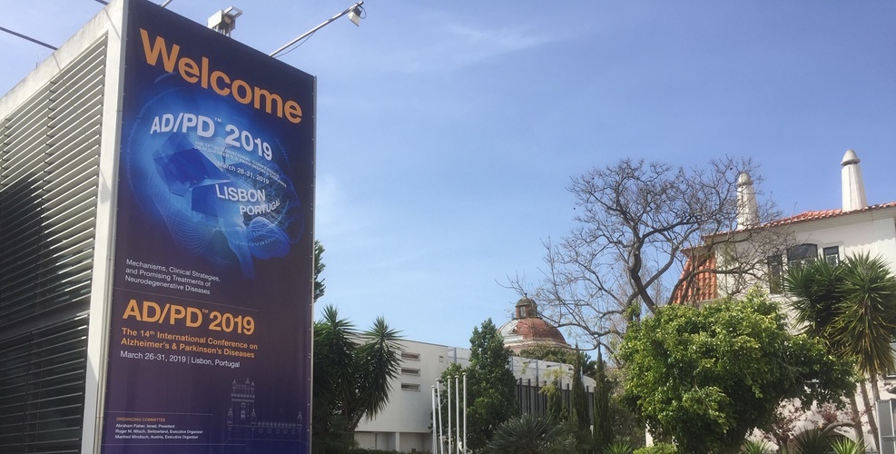
We don’t yet understand why this is the case but it must reflect differences in vulnerability, Glenda Halliday from Brain and Mind Center, University of Sydney, Australia, told a plenary session of ADPD 2019.
More glia than neurons accumulate α-synuclein. Immune-related receptors may be involved
Lewy bodies are only part of the pathology, and aberrant α-synuclein is not the only protein involved. But the fact that Lewy body inclusions are found in the majority of people with PD, and the fact that α-synuclein is the main constituent of their core fibrils, put it clearly in the frame. And we now start understanding reasonably well how Lewy bodies arise.
The process begins when α-synuclein forms punctate material. This comes together to form loose filaments and eventually becomes compacted. The average duration of the process may be ten or more years. It is accompanied by increased phosphorylation of α-synuclein, and an increase in the membrane-bound rather than the soluble form. Protein ubiquitination is present, but this does not lead to degradation. There is also the turning on of immune signals, such as the expression of Toll-Like Receptor 2 (TLR2), indicating an inflammatory response of the innate immune system.1
In multiple system atrophy (MSA), although clinical disease progresses at a faster pace, the formation of the characteristic oligodendrocytes cytoplasmic inclusions (CGIs) also occurs over a prolonged period.
It has not been widely recognized, but – in the synucleinopathies – it seems that more glia are involved than neurons, and they are involved from the outset. So glial function is likely to be important, and inflammation is increasingly seen as a crucial element in these diseases.
Lewy body formation is accompanied by dysfunction in lysosomes and autophagy
With time, more brain regions become involved. This is no longer controversial, though the sequence in which areas become affected may vary between synucleinopathies and be reflected in clinically different phenotypes even within PD. A limbic-dominant form, with spread via the olfactory bulb, has been proposed as an alternative to the classical Braak pattern of disease ascending from the brainstem.2
The Lewy bodies spreading was unequivocally demonstrated by the fact that embryonic dopamine neurons transplanted into the brains of people with PD develop Lewy pathology after around ten years.3 The pathology is probably passed cell to cell. We know that α-synuclein can be released from neurons by mechanisms such as exocytosis and through exosomes, but the mechanisms of uptake, and the amount that needs to be present to achieve transmission, remain unclear.
Whether spread is facilitated by an increase in vulnerability factors across the brain is also an open question. The possibility that glymphatic system dysfunction is involved in propagation deserves greater attention.
A further lesson from the brain is that forms of α-synuclein, particularly differences in solubility, really matter. They are different in MSA and PD; and we need to better understand the nature of the modifying factors.
Our correspondent’s highlights from the symposium are meant as a fair representation of the scientific content presented. The views and opinions expressed on this page do not necessarily reflect those of Lundbeck.