
Progress in Mind
Speaking at #WCN2021, Professor Todd Schwedt, of the Mayo Clinic, USA summarized that although there are clearly peripheral inputs, migraine is primarily a disorder of the brain. He presented insights into the accumulating evidence for this, which include studies of migraine symptoms, human neurophysiology, and imaging research.
The brainstem forms a functional unit with several hypothalamic nuclei capable of modulating diverse functions including migraine-relevant trigeminal pain processing, appetite, and arousal regulatory networks, and as such is a potential therapeutic target.1 (Holland 2019) While peripheral contributions in migraine stem from the trigeminal ganglion, trigeminal nerves, upper cervical nerves, and meninges.
Migraine symptoms localize to the brain
Migraine often presents with diverse premonitory symptoms, the majority of which localize to the brain, and include fatigue, cognitive dysfunction, neck stiffness, photosensitivity, phonosensitivity, irritability, speech difficulties, yawning, thirst, and urination. Symptoms associated with aura include visual, sensory, and motor inputs, and cortical spreading depolarization which may activate the trigeminal system.2
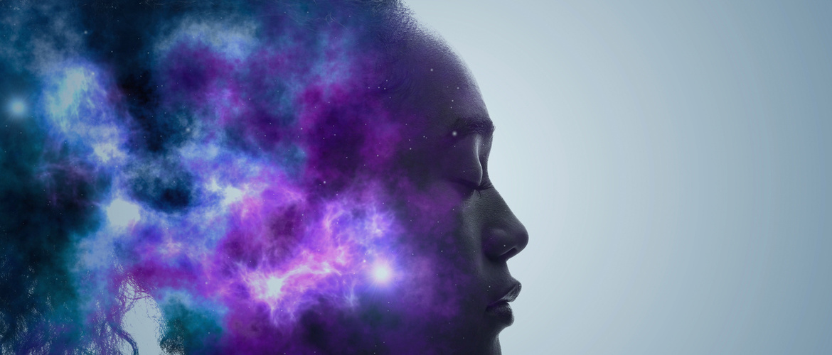
Sensory hypersensitivities are a key component of migraine
Sensory hypersensitivities are a key component of migraine and include visual, auditory, and olfactory stimuli, and cutaneous allodynia.3 Imaging studies of multisensory brain regions show that aberrant multisensory integration is involved in migraine.3 Multisensory integration is described as the brain’s ability to co-process and co-modulate different sensory stimuli to derive a unified perception of the environment.
The brain’s ability to co-process and co-modulate different sensory stimuli is negatively affected in migraine
Auditory, visual, and olfactory stimuli can trigger migraine attacks, and exposure to visual and auditory stimuli can worsen headache intensity. For example, neurophysiology studies show that visual stimuli impact trigeminal pain processing, and people with cutaneous allodynia are more sensitive to sound.4
Cortical information processing is abnormal between migraine attacks
Other neurophysiology studies show lower discomfort thresholds, a lack of habituation to sensory stimuli, central sensitization, and inadequate sensory inhibition in migraine. Response habituation during stimulus repetition – a fundamental, protective feature of cortical information processing – is abnormal in migraine between attacks.5 The abnormal information processing most likely occurs due to inadequate control by the state-setting pathways originating in the brain stem.5
People with migraine exhibit impaired conditioned pain modulation, suggesting a deficiency in inhibition of trigeminal nociception, which may contribute to the development of migraine headaches.6 Enhanced cognitive pain processing observed in brain imaging of people with migraine may reflect cerebral hypersensitivity related to high expectations and hypervigilance for pain. People with higher frequency migraine headaches have greater hyperactivity of pain facilitating brain regions.7
High frequency migraine headaches are associated with greater hyperactivity in pain facilitating brain regions
Localized as well as widespread increased mechanical sensitivity in cephalic and extra-cephalic regions is observed in all phases of the migraine cycle, which is even more pronounced immediately before, during and after a migraine attack.8 The spreading of multimodal allodynia and hyperalgesia beyond the location of migraine headache is mediated by sensitized thalamic neurons that process nociceptive information from the cranial meninges together with sensory information from the skin of the scalp, face, body, and limbs.9 While imaging studies show greater activation, less inhibition, and atypical structure correlating with symptom severity.2
Our correspondent’s highlights from the symposium are meant as a fair representation of the scientific content presented. The views and opinions expressed on this page do not necessarily reflect those of Lundbeck.